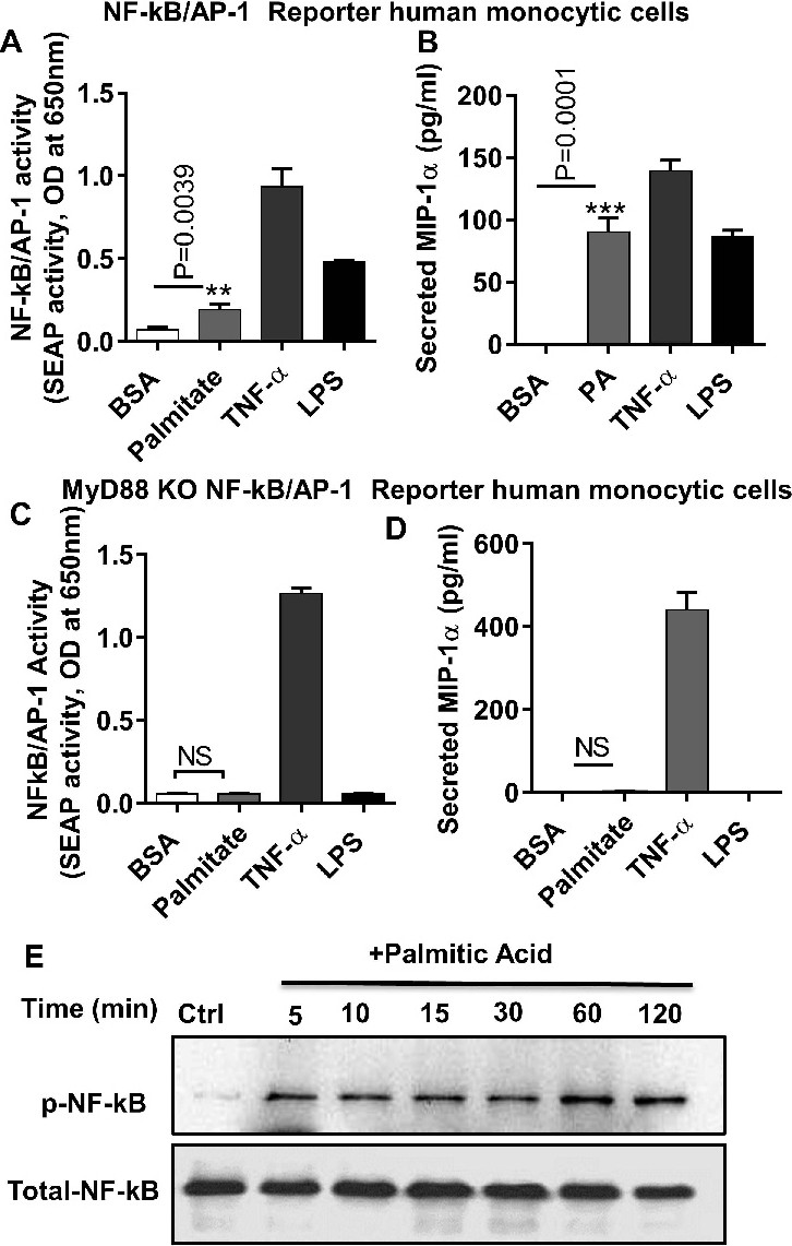
Fig. 6. NF-κB/AP-1 activation is associated with palmitate induced MIP-1α production. NF-κB and AP-1 reporter monocytic cells were treated with 0.1% BSA (vehicle) palmitate (100μM) or TNF-α (10ng/ml) or LPS (ligand for TLR4; 10ng/ml) for 24 hrs. Culture media were collected. Cell culture media were assayed for SEAP reporter activity (degree of NF-κB /AP-1 activation) along with MIP-1α protein production (A and B). NF-kB/AP-1 reporter MyD88 KO human monocytic cells were treated with 0.1% BSA or palmitate or TNF-α or LPS for 24 hrs. SEAP reporter activity (degree of NF-κB /AP-1 activation) along with the secreted MIP-1α protein was determined in the cell culture media (C and D). THP-1 cells were treated with palmitate for different time points and cell lysates were prepared as described in methods. Samples were run on denaturing gels. Phosphorylated NF-kB is shown in the upper panel and total respective protein is shown in the lower panel (E). The results obtained from three independent experiments are shown. The data are presented as mean ±SEM. Statistical analysis was done using t-test. P<0.05 was considered as statistically significant. **P< 0.001, ***P< 0.0001.